About the complex sex lives of hermaphrodite snails, who may or may not have a phallus: to be sung to the tune of ‘Oh dear, what can the matter be? Three old ladies stuck in the lavat’ry…’
(General chorus)
‘Dear, dear! What can the matter be?’
‘Oh dear! Two snails with aphally.’
‘See, dear – they’re hermaphroditidae.
Can they have fun at the Fair?’
(The aphallic snail puts its case.)
‘But I don’t need another, I’m father and mother.
No sexual behaviour’s an energy-saver.
A penis is silly, I don’t need a willy!
I have fun on my own at the fair.
(General chorus)
Hey ho! We’re Bulinus truncatus.
It’s so! Our sex-life’s the greatest.
Ho ho! Cross-breeders or self-maters,
We all can have fun at the Fair.
(The phallic couple’s case)
We’re both sisters and brothers, we’re like two paired lovers.
We each can inseminate, phallus can penetrate,
Not so! I am dominant, penis is prominent.
So which has most fun at the Fair?
(General chorus)
Ha ha! Our genetics are so complex.
Ooh ah! So too are our modes of sex.
Production of offspring is one of our main objects,
So our species can go to the Fair.
(The tetraploid’s lament)
With four sets of chromosomes, I’m host to schistosomes,
Humans would best avoid water near tetraploids.
A larva could bore in, through my unprotected skin –
Then I’d have no fun at the Fair.
(General Chorus)
Oh dear, this water’s too fast to drowse.
Help, dear, I just want to stay and browse.
Quick, dear! Stick your foot where the rock allows,
Or we’ll be washed away from the Fair.
(Exit)
(The survivor’s lament)
I’m male and I’m female, but I am a lonely snail —
No-one to talk or play, they’ve all been washed away.
I’m alive and I’ll thrive, and I hope my own eggs survive.
I’ll be joined by my kids at the Fair.
***
This piece of doggerel (it hardly qualifies as a poem!) comes from my novel Seaside Pleasures– and has some relevance to the story. One of the main characters is a malacologist/parasitologist …
The ‘singing snails’ are Bulinus truncatus, which are found in the shallow water at the edges of lakes in Africa.
Bulinus
These ‘tetraploid’ variants – they have 4 sets of chromosomes – are the intermediate hosts of an extremely debilitating disease in humans known as urinary bilharzia or schistosomiasis, caused by the parasitic worm Schistosoma haematobium. The male and female adult worms live in permanent copula in the blood vessels around the bladder; the female releases fertilised eggs, each of which has a spine on the egg-shell and gets pushed through the muscles of the bladder wall.
If the infected human pees into fresh water, the eggs hatch into tiny larvae, miracidia, which swim around until they find a snail; they burrow into the snail and undergo a complex multiplicative lifecycle inside it then burst out as another swimming stage, the cercariae, which can burrow in through a person’s skin. These migrate and undergo further development inside the human, finally ending up in the bladder wall.
***
A glass model made by the Blaschkas of Dresden, of the freshwater snail Lymnaea stagnalis (which, as you can see, is not aphallic)
When I was an undergraduate, I collected these green and red algae on a London university field trip at Liverpool’s former Marine Laboratory at Port Erin, Isle of Man. We were shown how to float the more delicate species onto white card; large polysaccharides like alginate and carrageenan would be released from the cells, swell as they hydrated, and bind the fronds onto the card as the specimen dried. After all these years the algae have kept their colour and shape. Larger, thicker algae like the brown Fucus species common on rocky shores, were pressed and dried.
Collecting seaweed and displaying it in albums was one of the ‘sea-shore pleasures’ that entertained the Victorian middle classes. Ladies were not expected to be scientific in their collecting but, rather, to arrange their seaweeds in an aesthetically pleasing and artistic manner; the making and displaying of scrapbooks was a parlour pursuit, art rather than science.
‘Seaweed and sentiment’, 1865
(Collecting sea-anemones – actinia – and keeping them in marine aquaria was another craze, inspired by Philip Henry Gosse’s Actinologia Britannica (1860): so much so that he was to bewail, ‘They have swept the shore clean as with a besom!’)
Amelia Griffiths (1768-1858), the widow of a clergyman, had become singularly knowledgeable about seaweeds. From her home-base in Torquay she collected along the Cornish, Dorset and Devon coasts. According to Philip Strange’s fascinating blog-post, “she helped male seaweed enthusiasts in producing scholarly studies on the larger and smaller seaweeds, generously giving her knowledge and donating samples.” Amongst the men was the Irish botanist William Henry Harvey (1811-1866) who went on – with Mrs Griffiths’ help – to produce his handbook of British Marine Algae, followed later by the 3-volume Phycologia Britannica, illustrated with coloured plates, and published in 1846.
Phycologia Britannica, title page
Delessaria hypoglossum, Plate 2
Plate 2, description
According to Philip Strange, Mrs Griffiths was often helped by her maid Mary Wyatt – who later ran a shop in Torquay selling shells and other seashore memorabilia to visitors, and who was encouraged by Harvey “to sell books of pressed and named seaweeds to help identification. Supervised by Amelia, she produced the first two volumes of Algae Danmonienses (Seaweeds of Devon) by 1833. Each volume contained 50 species …”
As an aside here, Philip Henry Gosse must surely have come across the elderly Mrs Griffiths (she lived to the age of 90) during his early sojourns on the Dorset and Devon coasts, during his own expeditions to collect and identify marine animals for his marine aquaria.
This was a time when science, in terms of enquiry, knowledge and new techniques, was advancing quickly. Scientists (‘natural philosophers’) knew of each other and, if they had been appointed members of prestigious establishment institutions such as the Royal Societies of Edinburgh or of London, they were sure to meet and talk and correspond.
At the same time as Harvey was working on the accurate representation of British seaweeds through coloured engravings, lengthy papers and postcripts and addenda were being presented to the Royal Society on other methods of capturing images, on paper or metal plates that had been made sensitive to light.
The oriel window at Lacock Abbey, an image of which Fox Talbot captured in a calotype
William Henry Fox Talbot was developing a method of recording images as negatives on high-quality paper sensitised with a solution of silver iodide, which became known as the ‘Talbotype’ or calotype process.
At the same time, the astronomer and chemist John Herschel (1792–1871), who – like many natural philosophers of the time – had wide and eclectic interests, had been testing the effect of the spectrum of light in changing the colours of plant extracts. He presented his findings to the Royal Society in a lengthy paper“On the Action of the Rays of the Solar Spectrum on Vegetable Colours and on Some New Photographic Processes,” on June 16 1842, with a postscript added in August.
As well as using extracts of various flowers, he also used a solution of the gum guaiacum, and experimented with the effect of chlorine gas and solutions of salts of chlorine, iron, ammonium and more.
Extract from John Herschel’s 1842 paper
He began to home in on the underlying chemistry and the best methods of producing – and fixing – the colour change of the pigments on exposure to sunlight, and to use this to produce negative images of objects placed upon the sensitised paper: a process he called cyanotype.
Extract from the postscript to Herschel’s 1842 paper
Rather than plant extracts, with their cocktail of constituents, he sensitised paper with a mixture of iron salts, ferric ammonium citrate and potassium ferrocyanate. Exposure to sunlight produced a background of ‘beautiful and pure celestial blue’ – Prussian Blue – around the object that was being recorded.
John Children was Keeper of the Department of Zoology at the British Museum from 1837-40, and was also a chemist and an entomologist. He too was a Fellow of the Royal Society and served as its Secretary from 1830-37. Naturally, he knew Fox Talbot and John Herschel; indeed, they were friends, and along with other scientists such as Humphrey Davy, visited his home – with its well-equipped laboratory – in Kent.
Children’s wife Hester had died soon after giving birth to their daughter Anna in 1799, and it seems that Anna later “received an unusually scientific education for a woman of her time”
In 1825 she married John Pelly Atkins, a London merchant, and they moved to Halstead Place, the Atkins family home in Sevenoaks.
Atkins was also acquainted with Fox Talbot, and so, through her husband’s and her father’s friends, Anna learnt about calotypes and cyanotypes, the new techniques of photography.
At the same time as William Harvey was being helped by Amelia Griffiths, Anna Atkins too began recording the variety of British seaweeds, but using Herschel’s photographic technique of ‘blueprinting’ or cyanotyping; she placed pressed algae under glass on paper that had been sensitised with a mixture of soluble iron salts, and exposed them to the sun. After washing in water, the background colour deepened to a uniform ‘celestial blue’, leaving the detailed outline of the algae in grey; washing, then drying, fixed the colour and the image. Since the object was placed directly onto the paper, these were strictly cyanograms.
Between 1841 and 1853, Anna produced the several parts of her Photographs of British Algae: Cyanotype Impressions, each of which contained around 400 images. It is not known how many copies she made but fewer than 20 still survive, all slightly different, and those at the British Library and the New York Public Library have been digitised and made available online. Inside the NYPL’s copy is the hand-written inscription, ‘Sir John F W Herschel, Bart. With Mrs Atkins’ compliments’. It is even possible to download a Kindle version (in shades of grey) from Amazon.
Title page
Laurentia pinnatifida
Delessaria sanguinea
Chondrus crispus
(All these images are from the New York Public Library’s digitised version)
Much of her work after 1853, such as the beautiful Cyanotypes of British and Foreign Ferns , was made in collaboration with her lifelong friend, Anne Dixon (1799-1864), a second cousin of the writer Jane Austen.
‘Bloom’
I belatedly came across Anna Atkins’ cyanotypes when my daughter Rachel gave me a copy of the Horniman Museum’s publication, Bloom, related to the exhibition by artist Edward Chell. In 2011 one of theHorniman’s librarians discovered that ‘in the darkness of the archives’ of the collections were folios of a rare and valuable copy of Atkins’ British Algae: Chell’s work during his residency at the Museum was inspired by the cyanotypes as a way or representing plants.
The technique
Atkins’ cyanotype ‘photographs’ are so striking because the background is so even in colour, and the outlines of even the most delicate algae are shown in such fine detail.
Channel wrack & spiral wrack on Parton beach
A mix of species in Allonby Bay
I already have a small collection of pressed seaweeds; I take people on low-tide walks on the Solway shore where we wander amongst weed-covered rocks; Rachel and I for several years developed and printed our own black-and-white photos; there is plenty of information about the cyanotype technique online (the Alternative Photography website is especially useful) … The technique is reputedly ‘simple’ – so of course I had to try it.
Basically, just two iron-containing chemicals are required: potassium ferricyanide and ferric ammonium citrate. Make them up separately – Solution A: 25 grams Ferric ammonium citrate (green) and 100 ml. water; Solution B: 10 grams Potassium ferricyanide and 100 ml. water – and mix in a non-metallic container.
Then – in dim light – paint the mixture onto paper using a soft, broad paintbrush (without a metal ferrule); leave the paper to dry in the dark; place your object or negative on the sensitised paper, preferably under glass to flatten and hold it in place, and expose to sunlight until you see the outline of the object.
First attempt: leaves and seed-heads
The pale green-yellow of the sensitised paper darkens towards blue in the sun, but is unchanged beneath the object. In bright sunlight the image develops within a few minutes but on a duller day it takes longer – and during a longer exposure the sun moves, so the outline of the object becomes fuzzy.
Wash in cold water for about 10 minutes until no greenish-yellow colour washes out (the startling Prussian Blue colour develops most strongly during washing) – and hang up to dry.
In the first step of the reaction, ferric (FeIII) ions from the soluble ferric ammonium citrate are reduced in a photochemical reaction, by UV in the sunlight, to ferrous (FeII). In the second stage, these ferrous ions then react in a complex way with the ferric ions in the potassium ferricyanate.
The result is an insoluble, dark blue compound – ferric ferricyanate – or Prussian Blue.
3Fe2+ + 2Fe(CN)63- → Fe3 (Fe(CN)6)2
After exposure, the colour is brought out further by oxidation during washing in tap water (dilute hydrogen peroxide can also be added).
Results
My first attempt, using leaves and seeds from the garden, worked astonishingly well, and I was thrilled with the colour; it was either ‘beginner’s luck’, or else the marine algae really are more difficult.
To make such fine and accurate prints as Anna Atkins’, with a uniform background colour, will require more experimentation, but I have been pleased with the results so far, and I’m enjoying the challenge. Each attempt shows me new variables that need to be controlled. Unfortunately I can’t control the amount of sunshine!
I’ve experimented with different papers, with different ways of coating them, with different ways of arranging the algae, and with different washing-times.
Careful arrangement of each specimen on the paper with forceps before lowering the glass is tricky – but important.
Shadows blur the edges
Partly-dried specimen refused to lie flat
I’ve learnt that thick seaweeds, even after pressing, may throw shadows that blur the edges. With very delicate algae, or colonial animals like hydroids, the finer details often appear during washing.
Art meets science: different points of view
Anna Atkins’ cyanotypes of marine algae and British ferns are a mixture of artistry and science, which she intended to be used as an aid to identification.
Mine cannot yet be used for accurate identification of species, but I’m enjoying the multifaceted approach: cyanograms, photographs of fresh specimens, pressed and dried specimens, and algae that have been floated onto card and dried.
Chondrus crispus, hydrozoan colonies, and red alga with mussel spat
A colonial hydroid, Sertularia
There is the possibility to play with different ways of recording shape and colour.
There is the possibility to gain different perspectives, and to hint at different points of view.
Just to reassure you, the novel is not ‘about’ embalming – the word is used as a metaphor for the many different ways in which we preserve our memories and ideas. Ruth is a taxidermist who lives in the northern Lake District in England – she is one of the three main characters. The others are Madeleine, a sheep-farmer (whose barn Ruth rents as a workshop), and Lisa (a mathematician and university academic from Liverpool). The paths of their lives, their loves, their interests, cross and interact.
Ruth’s blogs, placed at various stages in the narrative, contain clues to what might (or might not) have happened in the past.
There is more about the novel here: how to buy it (paperback or ebooks), reviews, illustrated sections on the Anatomists, patterns, achondroplasia, foot-and-mouth disease, taxidermy, some videos relating to ‘Lisa’, links to other extracts…
There are many ways of killing flies, but the extreme solution of first swallowing a spider is certain to cause discomfort.
Spiders – the non-bird-eating species – have evolved a variety of web-designs for snaring their prey. Much as a sea-trout swims into a net fixed across the mouth of a stream, so might a fly be caught, and held tightly by minute globules of glue. After a quick paralysing bite, the spider packages up the helpless creature in a silk winding-sheet. The sundew plant uses sticky globules too but its treatment is less aesthetic, the edges of its scarlet-fringed leaf curl over the insect, imprisoning it so that it stops wriggling and dies, and gradually, very gradually, its body is digested. All that remains on the web or in the sticky leaf is an empty husk, the insect’s scarcely recognisable, undigested exoskeleton.
Sundews wait with open leaves for unwary insects
Entomologists and butterfly collectors need their dead insects to be whole and unharmed. Gaseous carbon dioxide, freezing, fumes of chloroform or nail polish remover, or pickling in alcohol or formalin, the methods all have their advantages and drawbacks. To make a Killing Jar: place a layer of ten finely-chopped laurel leaves in the bottom of a honey-jar and screw the lid on tightly, to capture the marzipan fumes of hydrogen cyanide.
Kill and arrange aesthetically in your cabinet. Admire – and mourn. Cherish the memory.
Unlock and fold back the polished wooden bar on its bright hinges and slide out the shallow drawers, one by one. Look at the hundreds of beetles, thousands of green-eyed flies, their bodies pinned onto cork, the tiny labels with name and place and date. The ‘Type Specimen’, the acknowledged example of its kind, and dozens of variants with minute mutations and alterations. It’s like stamp-collecting, you must check the number of perforations, the graded colours of the patterns. But the modern collector does not merely tick the box but prefers to ask ‘how?’ and ‘why?’. The modern taxonomist deals in molecules not just in external signs, and seeks the remnants of genes in mummified or pickled corpses, or from the blood-meals of mosquitoes preserved in the sticky exudations of trees.
The Dutch anatomist and botanist Frederik Ruysch was a collector of insects as well as foetuses and curious natural objects. Insects symbolised the transiency of life on earth, the beautiful butterfly bursts forth from its papery brown pupa-case but lives for a few short weeks (unless it has the wit, or inbuilt programming, to hibernate). Adult Ephemeroptera, the mayflies, live for a single day, escaping one medium into another, from water to air, to fly and mate. Brief encounter, brief lives!
Ruysch’s moralistic and allegorical tableaux on the theme of death now exist only as engravings (drawn ‘from life’ by Huyberts) in the large leather-bound volumes of his Thesauri Anatomici, his Omnia Opera. Insects, bones and anatomical curiosites share the stage. The skeletons of foetuses stand or lie in different postures, amongst pebbles and kidney stones and taxidermal preparations of birds and other animals, arranged artistically on ornate wooden stands. In the tableau depicted in Thesaurus VIII, dried blood vessels project upwards as a forest of branches, a skeletal arm waves a scroll of paper, and two foetuses weep into lacy handkerchief of dried mesenteric tissue. Well might they weep, for the hollow rock beneath them is a dried injected uterus, cut and opened like the door of the Tomb to show the dead embryo inside. ‘A Fate, ah bitter fate!’ sings the foetal skeleton in Thesaurus III, as it accompanies itself on a malformed bone ‘violin’ with a dried artery for a bow. Its grinning companion encircles itself around with a snake of injected sheep gut, the vase-like form of an inflated testis adds artistry and elegance, and in the foreground a prostrate tiny skeleton holds an adult mayfly.
Title page, Ruysch’s Thesaurus Anatomicus
Explanation of the drawings by Cornelius Huyberts
From Thesaurus VIII
Her father’s collections provided a handy source of specimens for Rachel Ruysch. Moths and butterflies, grass-hoppers and flies rest amongst the richly-coloured, richly-textured flowers in her still-life paintings. A lizard, a frog, snails with striped shells – the invertebrate creatures have such depth and colour as to seem alive, halting briefly for a single moment in time. No other artist could capture the texture of velvety petals and reflections on the damp bloom on grapes so well as Rachel Ruysch.She became famous, her works were much sought-after, and in 1708, aged 44 years old, she was appointed Court Painter to the Elector Palatine of the Jülich-Berg duchy, Johann Wilhelm. She and her husband Juraien Pool stayed at the court in Düsseldorf until the Elector died.
An arrangement of flowers by a tree trunk, Rachel Ruysch (1664 – 1750). Painted in Amsterdam, where Ruysch spent most of her life, this reflects the Dutch mania for flowers. …The flowers move upwards in a spiral. The Dutch love of varied detail is seen in the insects, fungi, and lizard. The artist used a sponge to apply the paint to get the correct texture for the moss and pressed fabric against the wet surface to portray the scales of the lizard.
In Arrangement of flowers by a tree-trunk, in Glasgow’s Kelvingrove Museum, six different butterflies and moths fly amongst the flowers, one drifting down to alight on a pure white trumpet of convolvulus, another fluttering vainly between the flat jaws of a lizard. But these insects, unlike her father’s specimens, are not moralising statements for they exemplify reality, their details have been observed and recorded, and are offered to us in all their glorious detail.
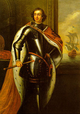
In 1697, Peter the Great, aged 25 years old, worked in an Amsterdam shipyard and attended Frederick Ruysch’s anatomy lectures. Peter enjoyed carving and joinery, he liked to work in wood and stone and human flesh, he even fancied himself as a bit of a dentist and a surgeon. His portrait was painted in Amsterdam by Jan Weenix, who later painted twelve large murals for the Elector Palatine. Perhaps Rachel sometimes stopped by to watch and advise: ‘Put a deer-fly or two on that stag’s skin.’
The portrait painter Godfried Schalken overlapped with Weenix in Dusseldorf in 1703 although not with Rachel Ruysch, but he had already painted her portrait in Amsterdam. In her late thirties she still has the same long straight nose but now she also has a double chin and lace at her bosom.
In 1699, a year after Peter left Amsterdam, boasting that he had bought his new boots with the proceeds of his work, Maria Sybilla Merian (1647-1717) visited Frederik Ruysch’s house to study his insect collection. She wanted to learn about insects, their metamorphoses and lives, and that same year she took her youngest daughter Dorothea and went abroad, to Surinam. She saw how caterpillars pupated into chrysalids from which a moth or butterfly crawled out, damp and crumpled, and spread its wings. She watched insects, she collected them, she wanted them alive as well as dead, and she painted their life-cycles and their food-plants.The details were as scientifically exact as the glass insects and flowers of the Blaschkas’ series on insect pollination. The colours glowed.
Thirty years previously the Leiden anatomist, Jan Swammerdam, famously dissected a preserved caterpillar for Cosimo de Medici. Swammerdam, according to the naturalist Boerhaave, wanted to preserve parts of the dissected body in constant readiness for anatomical demonstrations, freeing him ‘from the difficulty of obtaining fresh subjects, and the disagreeable necessity of inspecting such as were already putrified’. He, like all the other Leiden experimenters in embalming, used oil of turpentine (at that time, the distilled resin of the terebinthe tree, Pistacia): take a tin vessel large enough to hold the corpse or dissected organ, and place a grid in it two fingers’ width above the bottom; pour in oil of turpentine to three fingers’ width, place the corpse inside and cover the vessel tightly with a lid. An embryo requires two months, a spleen ten days, a liver one month. Swammerdam preserved the body of a month-old child – and a whole lamb.
The oil of turpentine converted the body fats of insects into a harder limey substance which could be washed away, leaving fibrous tissue intact. Inside Swammerdam’s dissected caterpillar de Medici saw the wings of the future butterfly, and saw too that metamorphosis was an unfolding of parts already present rather than an alchemical change. Swammerdam turned down de Medici’s offer of 12,000 guilders for his insect collection.
Houbraken’s engraving, taken from a portrait (dating from 1700) by George Gsell, portrays Sybilla Merian with shells and plants around her. She died in Amsterdam at the age of 70 and that same year, 1717, Peter the Great returned to Amsterdam and bought not only most of Frederik Ruysch’s great collection of tableaux, ‘naturalia’, embalmed babies and decorated foetuses, for 30,000 guilders, but also the late Sybille’sMetamorphosis Insectorum Surinamensis and part of her insect collection.
Maria Sybilla Merian, engraving from portrait by George Gsell
He also acquired Sybilla’s daughter Dorothea, persuading her to use her skills to design the decorations around his new collection in the Kunstkammer.
Gsell’s portrait of the giant Bourgeois
The skeleton of Bourgeois, in Peter the Great’s museum
Her husband (and Sybilla’s son-in-law) Georg Gsell, went too, and when the giant footman Bourgeois died in 1724, Georg painted him – posthumously – so that his fleshly appearance was preserved in a portrait before he was boiled.
Rachel Ruysch was long-lived like her father, and painted her last still-life at 83 years old, in 1749, less than a year before she died. In one of her paintings a caterpillar hangs by a thread. In Merian’s drawings from Surinam, caterpillars munch holes in leaves, spin silk around themselves to form protective cocoons, or their skin hardens and changes shape to form a pupal case or chrysalis. Inside the pupa, as Swammerdam had seen, organs and appendages shrink or grow, change shape, wings sprout, and antennae.
But now we know that parasitic wasps may lay their eggs in living caterpillars, eggs which hatch into larvae that devour the caterpillar’s tissues from inside, the wasp larvae growing, moulting, themselves changing shape until they burst out, leaving an empty husk behind them. Did Merian and her daughter observe this metamorphosis of aliens, this apparent transmutation of species? The caterpillar as host, the food-provider, the life-support system, the container for a new life-form.
So also some modern matryoshki, variants of the wooden nested Russian dolls. The traditional dolls with rosy cheeks and peasant scarves, one inside another, date from the late 1800s, long after Peter the Great practised his own wood-turning skills. The mother figure is a symbol of birth and renewal, the handing on of the lineage and the mitochondrial genes. But modern versions of the matryoshka escape the human form, so that a cow contains a sheep, which contains a pig, a chicken and, of course, an egg. The outer form hides its inner secret wherein one species generates another.
The recipe for an Elizabethan dinner does this too, but in reverse for it symbolises death rather than birth. It omits the mammals and sticks with our feathered friends. ‘Take a boned swan, stuffed with a boned goose, stuffed with a boned capon, stuffed with a boned muscovy, followed by a boned pheasant, then a partridge, a woodcock and a dove, stuffed with a boned snipe, a boned sparrow and a dozen lark’s tongues.’
As for that old lady who swallowed a fly, she took in the whole range from mammals to invertebrates:
There was an old lady who swallowed a cow.
I don’t know how she swallowed a cow!
She swallowed the cow to catch the sheep…
She swallowed the sheep to catch the dog…
She swallowed the dog to catch the cat…
She swallowed the cat to catch the bird …
She swallowed the bird to catch the spider
That wriggled and jiggled and wiggled inside her.
She swallowed the spider to catch the fly.
But I dunno why she swallowed that fly
Perhaps she’ll die.
And as for the old lady who swallowed a horse, what did you expect? ‘She’s dead, of course.’
Louis van Bils sold his secret recipe for embalming bodies to the States of Brabant and the University of Louvain, in 1664; it required copious quantities of rum and brandy. In 1669 de Bils also tried to sell his recipe in the northern Netherlands, in Leiden and Amsterdam, with the help of Dr Tobias Andreae, ‘Professor of Physick at Duysburgh on Rhyn’, but they met considerable resistance from Dr Frederik Ruysch. His own recipe was secret, after all, and would later be the envy of a Russian Czar.
These days embalming is a quicker, cheaper process, syringes attached to electric pumps, cheap ready-made preservatives, and suction tubes. Let’s not bother with the details. Better to drink the rum and brandy with the funeral meats.
Regnier de Graaf, 17th-century Dutch anatomist and physician (and also a Fellow of the Royal Society in London) whose name lives on today in the Graafian follicles of the ovary, devised a syringe that Jan Swammerdam found very useful. By then, the European anatomists were all at it, shooting up warm coloured waxes or mercury or resins into blood vessels and lymphatics, and dissecting tissues or dissolving them away – corrosion casting – to follow the course of the now-visible vessels. (Some still do it today, with the modern techniques of injection and plastination. Anatomy can no longer keep a secret.)
William Hunter, surgeon, accomplished teacher and accoucheur to royalty, and his brother John, surgeon and anatomist, their assistants and their protegés – they were all busy injecting and dissecting, delving into the twists and anastomoses of the body’s vessels, in chickens and alligators, rabbits and frogs, humans, dogs and leopards. For how could one perform corrective surgery on humans in the metaphorical dark? Comparative anatomy was a useful tool, for what might be indistinct in one species might be more clearly visible and understood in another.
De Graaf started with the proverbially fecund rabbit, William Hunter worked with women. In 1751 he procured the body of a woman in late pregnancy, and during the next 20 years he studied pregnancies at various stages. There are ten life-size plaster models of the gravid uterus in Glasgow University’s Hunterian Museum, each uterus in situ in the abdomen, the mother’s thighs sawn through to expose gigots of meat. No lacy caps disguise the stumps, this is anatomy for instruction not moralisation. We don’t know whether the real-life mothers were prostitutes nor do we care. The colours of the plaster casts are as fresh and bright as though uterus and foetus are alive, pink flesh, red and blue blood. The complex spatial puzzle, the three-dimensional fit between mother and child is made clear. ‘The whole of them are exactly like nature herself, and almost as good as the fresh subject’.
from William Hunter’s “Anatomy of the Human Gravid Uterus, Jan van Rymsdyck 1774
Ivory and glass eye, photo by Rosamond Wolff Purcell
An arrangement of flowers by a tree trunk, Rachel Ruysch(1664 – 1750, Dutch)Netherlands, Amsterdam (place of manufacture)oil on canvas36 7/8 x 28 in; 93.7 x 71.1 cm; framed: 1186 x 865 x 130 mm Painting entitled ‘An arrangement of flowers by a tree trunk’, by Rachel Ruysch. Painted in Amsterdam, where Ruysch spent most of her life, this reflects the Dutch mania for flowers. Such pictures found a ready market with rich and noble patrons. It is an early work of the type known as ‘forest floor piece’. The flowers move upwards in a spiral. The Dutch love of varied detail is seen in the insects, fungi, and lizard. The artist used a sponge to apply the paint to get the correct texture for the moss and pressed fabric against the wet surface to portray the scales of the lizard.45
(See below (1) For further information on these images)
Hunter employed artists like Jan van Rymsdyck to draw and paint pictures of the injected and dissected specimens at each stage. The texture of the life-size paintings in The Anatomy of the Gravid Uterus (1774) is almost the equal of Rachel Ruysch’s skills, a window is reflected in the damp membrane around a life-size foetus rather than on the moist bloom of a Ruyschian grape. Or on the glass cornea of an artificial eye.
Amongst the many specimens in the Hunterian Museum is a glass receptacle containing one of William’s injected placentas, a grey slab of tissue through which the silvery worms of mercury coil, anastomose and interdigitate. Who discovered what? John Hunter, too, had worked on the human uterus. Dispute arose between the brothers, and Benjamin Franklin again poured oil on their troubled waters. In Joshua Reynold’s portrait of John, the glass jar standing close to Giant O’Brian’s bony feet holds a section of placenta injected with red wax.
John, now Surgeon-General in the army, experimented on bringing dead bodies to life, or the near-dead, anyway, bodies rescued from near-drowning. By pumping warm air into their lungs, they were resuscitated and began to breathe again. Although he dissected and described the electric organs of the ray Torpedo, he did not try to use electricity to galvanise bodies to life. That was for Mary Shelley to imagine. But in 1818 James Jeffroy, Regius Professor of Anatomy at Glasgow, used bellows to blow air into the lungs of the dead murderer Matthew Clydesdale. The terminals of the newly-invented galvanic battery had already been connected to Clydesdale’s body, and when the current was switched on, his eyes opened, his tongue protruded and his lips moved. Did Jeffroy believe the man had come back to life? He plunged a scalpel into the murderer’s carotid artery, and the man ‘fell dead on the floor’. Onlookers fainted!
Dr William Hunter and his colleague Dr William Cruikshank put the injection technique to a less honourable use. In 1775 they embalmed Mrs Mary van Butchell, aged 36 years, with the help of her dentist husband Martin. Hunter’s recipe was quick and simple. Mix 5 pints oil of turpentine, 1 pint Venice turpentine, 2 fluid ounces oil of lavender, and vermilion. The solution was injected into Mary’s blood vessels until all her skin took up the reddish colour and the body was left to lie for a couple of hours to allow the solution to diffuse further through her tissues. Then her viscera were removed and washed in water, and injected and steeped in camphorated ‘spirits of wine’. She was injected a second time, her viscera were replaced and the body cavities filled with camphor, nitre (potassium nitrate) and resin before being sewn up. The ‘outlets’ of her body were also filled with camphor and her skin was rubbed with oils of rosemary and lavender. Wearing a lace dress and with a rosy-pink complexion, Mary was laid on a bed of Plaster of Paris to absorb moisture and, it is said, was displayed in the window of Martin’s house.
It is also said that this bizarre display was (a) to drum up custom for Martin’s dental practice and (b) because Martin could have access to Mary’s money only while she was ‘above ground’. Whatever version of the truth, the second Mrs van Butchell was unimpressed. Mary was sent to stay at Hunter’s museum in Great Windmill Street.
Preservation for the after-life, preservation as an object of worship, preservation to ‘make a point’, icons and auto-icons, anatomical aids and anatomical art: we humans can rationalise almost anything that we do. Lenin, a modest man, did not have much say about the matter after his death, but Jeremy Bentham (died 1832) definitely wanted to hang around and be useful. It’s a myth that he wanted to be displayed at University College London, for he wished to be useful as an anatomical specimen, and it’s a myth that he was embalmed. He was a taxidermal preparation, dressed in his own clothes which Professor George Thane noted in 1898 ‘were stuffed with hay and tow around the skeleton, which had been macerated and skilfully articulated.’ The head of the specimen is ‘so perfect that it seems as if alive’ – and is made of wax. Bentham’s real head was mummified and dried, not unlike one of the shrunken heads in Oxford’s Pitt Rivers Collection. Wrapped in a bituminous cloth, it was originally placed out of sight inside his torso.
The dead have so many secrets, so many different stories.
Frank Buckland did not mention the state of his hero John Hunter’s body when he found the coffin in St Martin’s vault but there are instances of bodies that fail to decay. Some of the 18th – and 19th-century bodies excavated from Christ Church crypt in Spitalfields in 1984 were preserved by saponification, their fatty tissue having converted into a stable brownish-white wax of saturated fatty acids and their salts This adipocere characterises the famous ‘Soap Lady’ whom Joseph Leidy gave to Philadelphia’s Mütter Museum in 1874. Perhaps the same had happened to Dante Gabriel Rossetti’s wife, Elizabeth Siddall. It is said that she remained intact and beautiful when the impoverished drug-addicted poet sent Charles Augustus Howell to retrieve his poems from her coffin (though it’s certainly a myth that her red-gold hair had continued to grow so as to fill the coffin).
Not so beautiful are the bog-bodies nor are they as well preserved as the fatty ‘bog-butter’, 2000-year-old wrapped packages recently found buried in an Irish bog. But the torso of Lindow Man from a Cheshire bog and the bodies of Tollund and Grauballe Men from Denmark are a couple of thousand years old, and probably the victims of ritual killing. Tanned, shrunken but preserved by the anoxic acidity of peat, with teeth and hair intact, they can still reveal some secrets and Grauballe Man is the most revelatory of all.
Dissections are out of the question because these bodies are valuable archaeological specimens, but computerised tomography or CT scanning uses X-rays to generate 3D images so we can nevertheless look inside. Grauballe Man doesn’t have a hard skeleton because the acid peat has dissolved the calcium, so his ‘bones’ are more like rubber. And his face has been re-modelled by the earlier conservators at the museum who padded the skin under his eyes with putty like a Pharaonic mummy. Sadly for Irish Cloneycavan Man, his face has been re-modelled too, but by the murder weapon. But he still has lovely red hair, slicked into a Tin-Tin quiff with Iron Age hair-gel, a mixture of vegetable oil and the resin of a French or Spanish pine.
No scalpel is needed now to look at bones and sinews, no syringe to find blood vessels and lymphatics. Sophisticated machines can look inside our living bodies, we can even watch our heart beat. We can even watch our foetuses suck their thumbs.Twin CT-scanners, combined with positron emission tomography, PET, can create pictures of the insides of living bodies as they have never been seen before.
As for the preserved dead, we can analyse the DNA of their last meal. ‘Ötzi’, deep-frozen for 5000 years in an Ötztal glacier, ate ibex and red deer and vegetables before the arrow penetrated his shoulder.
Mis-quoting Papin, ‘Through thy science … a dead person lives and teaches and, though speechless, still speaks’. But the story, mis-remembered, can speak with many tongues.
***
(1) Reflection of a dissection room window in the chorion (outermost membrane) covering a foetus – for more on Jan van Rymsdyck’s drawings and etchings of William Hunter’s dissections of the gravid uterus, see The Sterile Eye blog.
The artificial eye is in Peter the Great’s collection in St Petersburg and was photographed by Rosamond Wolff Purcell for the chapter ‘Dutch Treat: Peter the Great and Frederik Ruysch‘ in Finders, Keepers, Stephen J Gould and Rosamond Wolff Purcell, Hutchinson 1992 (she generously gave Ann Lingard permission to use the image on the cover of the First Edition of The Embalmer’s Book of Recipes). There is a short video about the book on YouTube.
Rachel Ruysch’s painting, An arrangement of flowers by a tree-trunk, is in Glasgow’s Kelvingrove Art Gallery and Museum. (Ann Lingard thanks the ‘Culture & Sport Glasgow (Museums)’ for permission to use this image.)
Glass sea-anemones, based on PH Gosse’s engravings, by the Blaschkas
Glass eyes, by the BlaschkasSierra Exif JPEG
White spheres lie on cotton-wool in the compartments of a flat wooden box: blue, brown or green circles, dark centres – pot-boilers, money-spinners – artificial human eyes.
On a workbench there are small bottles, pin-boxes and wooden trays containing different body-parts: glass tentacles of different shapes and colours, sponge spicules, tiny shells. ‘Mix ’n match’ invertebrates amongst the powdered glass and pigments. In 1860, Philip Henry Gosse’s Actinologia Britannica was published, illustrated with engravings of coloured sea-anemones and corals. A few years later, an Englishman living in Dresden showed these pictures to glass-maker Leopold Blaschka. ‘Marine creatures preserved in spirits look like grey rubber,’ he said. ‘Why not show their true colours by modelling them in glass?’ Leopold accepted the challenge: he had already made glass orchids, so now he merged his art with science, modelling the exquisite and minute details of invertebrate animals, making them objects for the museum and scientific study rather than ornaments for the drawing-room.
Later, he and his son Rudolf were to give up these animals and Haeckelian embryos for a ten-year commission to make the Harvard glass flowers, but in those early days their business was not yet profitable – so they created jewellery, and glass eyes for cosmetic use by the blind.
We don’t know where Frederik Ruysch obtained the glass eyes to fill the orbits of his embalmed and Death-defying Dutch babies in the 17th century but they obviously did the trick because the babies’ winning looks won the heart of great Czar Peter.
‘Auto-icon’ of Jeremy Bentham, University College London
The philosopher Jeremy Bentham, who died in 1832, probably had the foresight to choose his own glass eyes (but he clearly had not planned that his mummified head would be stored in the dark inside his torso).
Can we defy Death, and preserve and repair our ageing body-parts, can the blind really be made to see again? Human eyes are such complicated balls of cells.
William Paley had argued in his Natural Theology in 1802 that the eye, like a telescope, could only have been designed by a Maker. (But the Maker must have been having a visual migraine when he designed the mammalian retina, back-to-front.)
Charles Darwin struggled to understand how these ‘organs of extreme perfection’ could have arisen from chance mutations alone. ‘To suppose that the eye … could have been formed by natural selection seems, I freely confess, absurd in the highest degree,’ he wrote. It’s almost enough to turn one into a Creationist or propose the intelligent mind and hand of a Designer. There is the implication that Evolution had foresight and saw its future goal.
We know now that Evolution is a conservationist and throws very little away: ‘You want an image-forming retina? There’s a bit of photosensitive pigment kicking around somewhere. A bit of this and a little bit of that, let’s try them in this order instead …’ The ingredients are mixed in a different sequence, to a different recipe.
The ingredient pax6 has never been allowed to rest, we need that gene as much as a flatworm does. In the embryo of a fly the product of pax6 directs the development of the eye. Gosse examined an insect’s eye under his microscope in the 1850s: ‘How gorgeously beautiful are these two great hemispheres that almost compose the [dragonfly’s] head, each shining with a soft satiny lustre of azure hue,’ he wrote. ‘You see an infinite number of hexagons, of the most accurate symmetry and regularity of arrangement.’ Each of those ‘hexagons’ contains a lens, and a careful anatomist with a steady hand can prepare an insect’s eye so as to look through it himself.
Van Leeuwenhoek (who lived a mere 50 years from 1675-1725, but achieved so much), looked through his microscope fitted with the prepared eye of a honey bee at a church steeple (‘which was 299 feet high, and 750 feet distant’) and saw multiple inverted images of the steeple. It scarcely seems possible that van Leeuwenhoek could have seen and understood so much of the natural world through a handheld instrument – the microscope is displayed in the Boerhaave Museum in Leiden – that is barely two inches tall.
The surface of the fruitfly Drosophila’s eye resembles a small raspberry. If the pax6 gene is injected into the embryonic cells that should form a Drosophila leg, the raspberry-like eye grows instead. Even more exciting and astonishing is that the raspberry will also grow if the pax6 equivalent from a mouse is injected into the fly embryo! (1) A mouse’s eye is like ours, as different from a fly’s eye as is a fly’s wing from a bat’s. And so it is with pax6 from the squid which, like the octopus, is almost unbelievably related to creeping slugs and snails, and which has an eye that is superficially like a mammal’s (this time, God designed the retina the right way round). Squids, jet-propelled and predatory, need their big eyes to see their prey – the Blaschkas’ glass squid is translucent and delicate, with lustrous dark eyes.
The pax6 gene in the living, growing animals has been conserved, and put to different uses. Biologists have identified it and its related ingredients, they even know something of the recipe from which a human eye is made, but they cannot reproduce it in a culture-dish, they cannot make eyes. Yet.
Biologists can ‘make’ different sorts of cells. Take a fertilised mouse or human egg and nurture it in a culture dish for several days so that its cells divide and divide again, to form a hollow blastocyst. Imagine a football with a pork-pie suspended inside it, and shrink it down in your imagination to the size of a pinhead: that is a 6-day blastocyst and the ‘pork-pie’ is a ball of embryonic stem cells. Stem cells, that each contain the complete book of recipes to form any other type of cell in the growing embryo; ‘the secret of eternal life’, a cellular equivalent of a perpetual motion machine, the magic ingredient that will allow us to repair ourselves for ever. Not quite, but stem cells have their uses.
Van Leeuwenhoek looked through his tiny microscope and watched red blood cells circulating in the blood-vessels of a tadpole’s tail. He would have thought it almost unbelievable that stem cells from a blastocyst could be turned into red blood cells in a culture dish; or into nerve cells, or muscle cells. (He would have been even more disbelieving to see how frogs’ eggs could be manipulated to produce clones).
Scientists can persuade corneal stem cells to grow new pieces of cornea, they can even persuade embryonic stem cells to change into a kind of retinal cell. But they cannot yet grow an eye, and if they could, how would they rewire it to the brain? All those millions of wires bound together in a cable, each needing their own connections in the brain. The Designer made an unintelligent muddle with those wiring diagrams, too, crossing over the cable from the right eye’s socket to the left brain, and vice versa.
William Hunter, FRS (1716-1783), anatomist, and man-midwife to Queen Charlotte and the gentry, dissected many corpses throughout his studies and demonstrations of anatomy. As President of the Royal Academy, he also stressed the links between science and art, and commissioned paintings and drawings of the three-dimensional structures that he dissected, as aids to surgery and deconstruction. Many of the contents of his London collection were transferred to Glasgow after his death. Upstairs in Glasgow’s Hunterian Museum, on a wooden shelf, are multiple rows of jars containing eyes. Intact, they stare at you while you stare at them and, because they are dissociated from their faces you cannot tell whether they stare in hatred or fear or even, perhaps, amusement. Why did Hunter collect so many? Was he working on a study of the anatomy and development of the eye?
Clemente Susini: Organ of Sight. Wax model, 1803
But look at this. A woman sleeps sweetly, unaware that her body has vanished leaving only her head with its soft pink lips and perfect teeth, nestling on a silken cloth. We need only see her head because Organ of sight shows the blood and nerve connections to the eye, as known in 1803. Clemente Susini’s wax woman’s eyes are closed, so we need not be afraid.
But the eyes of Ana Maria Pacheco’s wooden sculpted heads engage you at once – stark white with dark irises and pupils, outlined by thin black lines, they look out at us slyly, laughingly, wryly, openly, in sadness and in pain, from their painted wooden faces. In Dark Night of the Soul (1999) the naked victim has his head covered by a cloth so that he may not see the archers aiming at him, or perhaps that we may not look into his eyes and see the terror in this so-called terrorist’s soul. The eyes of the carved watchers reveal their despair and make us weep.
Detail from ‘Dark Night of the Soul’, 1999; polychromed wood and gold leaf.
Elsewhere, thirteen heads stare out of a compartmentalised box (Box of Heads, 1983) and their white faces and gashed red mouths catch our glance one by one, and hold it. Individuals, their thoughts are almost visible and we are forced to wonder about them, for though disembodied they seem all too real. (It is not just their eyes, but also their teeth, that make them real, the porcelain teeth, with all the irregularities and imperfections of individual human mouths). The tiny boxed heads are as pale as bleached skulls.
In a tall wooden Cabinet in Museum Vrolik, Amsterdam, rows of skulls are supported by pegs on ebony stands. Hydrocephalus, microcephalus, bathrocephalus, they are all sizes, distorted, grinning toothily but without eyes. Shelves of skulls, a presentiment of the Killing Fields. Nearby is the Curator’s ‘favourite’ specimen, a little foetus with a fuzz of pale red hair, his arms hanging gently in the liquid preserving liquid as though he is merely resting. He is a little ‘cyclops’, whose genetic recipe made for him only one small central eye. He did not live to see the light of day, nor have the good fortune to enter the Country of the Blind.
***
(1) In the mid-1990s, Walter Gehring and his colleagues performed a series of what turned out to be ground-breaking experiments in the field of developmental genetics – they showed that the gene pax6 which initiates the early stages of eye-formation in the mouse could also initiate eye-formation in Drosophila. Not a mouse eye – but a fly eye. In other words, if the mouse gene was inserted into fly cells that would normally form a leg or antenna – a fly eye grew instead! See Figures 3 and 5 of Induction of Ectopic Eyes by Targeted Expression of the eyeless Gene in Drosophila
This glass and ivory eye in Peter the Great’s collection in St Petersburg was photographed by Rosamond Wolff Purcell, who very generously allowed the author to use the image on the cover of this First Edition
On July 5th 1893 an Exhibition of ‘A Collection of Hunterian Relics’ opened at the Royal College of Surgeons, in London. Not the relics of Dr William Hunter, FRS and President of the Royal Academy, but of his younger brother, John, the naturalist and surgeon, born in Lanarkshire in 1729, and died, very suddenly, in London on October 16th 1793.
Amongst the items on display is a ‘Copper, in which the Irish Giant, Charles Byrne, who was exhibited in London as O’Brien the Irish Giant, was boiled.’ The copper was lent by Professor Chiene, of Edinburgh. The catalogue has a further explanatory note: ‘This copper was in Hunter’s house in Earl’s Court and was sold there in 1866 when the house was pulled down. On the death of Byrne in 1783, Hunter obtained his body and macerated it in this copper … The skeleton of Byrne is in the College Museum.’ As indeed it is, to this day.
As for poor Hunter’s sudden death, there is also exhibited the ‘Sofa on which John Hunter died. Whilst speaking at a meeting of the Board of Governors at St George’s Hospital on October 16th 1793, Hunter was contradicted by one of his colleagues. He immediately left off his speech and in an excited state hurried to an adjoining room; where he fell into the arms of Dr Robertson and almost immediately expired.’ Fortunately for him, his body was not macerated in the copper, but was placed in a vault at St Martin’s in the Fields. Of which, more later.
The College Museum has an engraving of Sir Joshua Reynolds’ portrait of Hunter, and Byrne is thus twice immortalised for, in the top right corner of the portrait and behind Hunter’s head are the lower portions of two gigantic femora and their two feet – the legs of that most famous Tall Man who, from the angle of his skeletal feet, must be standing on tip-toe, perhaps to increase his height even more.
Portrait of John Hunter, miniature copy by Henry Bone RA after Joshua Reynolds, 1798. (My thanks to The Hunterian Museum at The Royal College of Surgeons of England for permission to reproduce this image.)
John Hunter has a square, kindly, thoughtful face and his forehead is broad and tall beneath his curly reddish hair. He doesn’t look like a giant-killer.
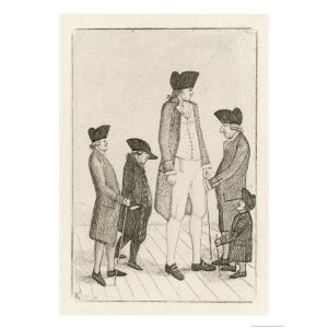
To be fair, it seems that poor Charles Byrne, aged 22, height 7 feet 7 inches, drank himself to death. The story of Byrne is well-known, much has been written about him, both fact and fiction. He was born on the Derry/Tyrone border in 1761 and, because those were times when the exhibition of ‘freaks’ was commonplace, Jack Vance, from a neighbouring village, persuaded him that he could get rich if he toured the shows and fairgrounds: a sideshow attraction. On April 11th 1782, Byrne and Vance (self-important as the Giant’s Agent) reached London and on April 24th an advertisement proclaimed ‘Irish Giant. To be seen this and every day this week, in his large elegant room, at the cane shop, next door to late Cox’s Museum, Spring Gardens.’ Admission was half-a-crown. By the late autumn, the public was jaded, the novelty had worn off, the price had dropped and the Giant O’Brien had moved twice, to rooms no longer so large and elegant. A year after his arrival in London, he was robbed outside a pub – of £700, presumably his entire worldly wealth which, for some reason, he was carrying in his pocket.
Despair, too much alcohol, and ill-health – Charles Byrne knew he was dying and somehow he also learnt that the anatomist John Hunter was very keen to obtain a giant’s skeleton. Byrne didn’t want his bones to be boiled, he didn’t want to be exhibited any more, alive or dead. He gave instructions that after his death his body should be watched day and night until a large lead coffin had been built. His body was to be placed in it and it should be carried (how many tonnes would a giant lead coffin weigh?) out to sea and sunk. A newspaper reported after the event that ‘Byrne’s body was shipped on board a vessel in the river last night … to be sunk in 20 fathom water: the body-hunters … have provided a pair of diving bells, with which they flatter themselves they shall be able to weigh hulk gigantic from its watery grave.
No diving bell was needed. Hunter, having sent his assistant Howison as spy, bribed the men whom Byrne had employed to sink him. Fifty pounds, one hundred pounds, the price escalated. Five hundred pounds was later reported as the sum involved. Byrne’s body was removed and carried by hackney coach then Hunter’s carriage to Earl’s Court where, since Hunter feared that his body-snatching would be discovered, Byrne’s body was ‘quickly cut to pieces and the flesh separated by boiling.
‘I lately got a tall man’, Hunter wrote to Sir Joseph Banks.
Presumably John Hunter and Howison (not to be confused with his brother William Hunter’s assistant, William Hewson) boiled and re-assembled the skeleton themselves. Ten years earlier William Hunter had fallen out with Hewson, for Hewson ‘had employ’d a Man to pick Bones out of the Tubs and fit up a Skeleton for him, without Leave’. Benjamin Franklin was called in to settle the dispute (Benjamin was often called in for this purpose). Although Hewson was highly skilled at injecting lymphatics and preparing specimens, he was eventually dismissed – and disinherited.
TH Shepherd’s engraving of the Museum at the Royal College of Surgeons, London
In Shepherd’s 1840 engraving of the College Museum, Charles Byrne’s skeleton with its smiling skull towers over the other exhibits from the top of a mahogany case. A photograph taken in 1852 shows him prominently displayed.
Brendan Holland and Marta Kobonits examine Charles Byrne’s skeleton in 2011 (see note 1)
And so he remains, in the 21st Century, and we think of him kindly and with sympathy. Perhaps finally he has been able to smile at the thought that his story as well as his bones (and a portrait of his feet!) are still preserved and admired nearly 250 years later.
The copper with which Charles Byrne is unfortunately associated was lent to the Exhibition of Hunterian Relics by a Scottish professor; Frank Buckland, Esquire, son of Dean Buckland of Islip, Oxfordshire, lent a Chair. The chair bore a brass plate with the following inscription: ‘This Chair is made from the bedstead of John Hunter…’ Buckland had been given the bedstead by ‘Professor Owen, FRS., etc. who wrote “ … it is the frame of the bedstead in which John Hunter lay when brought from St George’s Hospital”. ’
What a tangled network of old boys’ ties! Owen, of dinosaurs and founder of the Natural History Museum, the devious Owen, a curator of the Hunterian Collection – and Frank Buckland (1826-1880), naturalist, collector, taxidermist, expert on fish and fisheries, and a kindly, well-liked man.
Nearly 100 hundred years after the giant Byrne’s death, in May 1871, Buckland was visited by ‘a strange party from the other side of the Atlantic’: Miss Swan the giantess (7 feet 6 inches) and Captain Martin van Buren Bates, ‘about as tall’, and both aged 24 years old. Bates was ‘a splendid-looking fellow, very unlike pictures of the giant in the “fe fa fum; I smell the blood of an Englishman” legend.’ Not only was Bates splendid, but he and Miss Swan apparently made a splendid couple, too. ‘I make bold to say that Miss Swan is the most agreeable, good-looking giantess that I have ever met,’ Buckland wrote. ‘She is ladylike in manners and address and would be a most agreeable neighbour at a dinner-party.’ He had the opportunity to test this three years later, when he entertained the splendid couple at dinner in honour of their marriage.
Giants came to Britain from all around the world, Chinese, American, Irish and French. Buckland dined with ‘the Chinese Giant’, Chang Woo Gow. Buckland himself was the cause of a ‘breach of discipline’ in his regiment, a spreading roar of laughter one Sunday in 1862, when he appeared on church parade in the company of ‘Brice the French Giant and a dwarf then exhibiting in London.’ Brice, like the skeletal Byrne, was apparently 7 feet 7 inches tall, and a well-proportioned and amiable man. A frequent visitor to Buckland’s house, he gave him a pair of his shoes and a cast of his hand as mementoes. There is a story that ‘A lady dwarf was one day invited to meet him, but with untoward results; the good-natured giant took her up, as a little girl, on his knee, causing an explosion of indignation. “I am nineteen,” she cried, “and to treat me like a baby!” It was long before her ruffled dignity could be appeased.’
As for John Hunter, his body was deposited in the vaults of St Martin’s in the Fields in 1793. In 1859 Frank Buckland determined to find the body of his hero, ‘the greatest of Englishmen’, and spent two weeks searching through the vaults. ‘The stink awful’, he wrote in his diary. Then on February 22nd he ‘found the coffin of John Hunter. At work all the morning and about three in the afternoon found it, the bottom coffin of the last tier but one. It is in excellent condition, and the letters on the brass plate as perfect as the day they were engraved. “John Hunter Esq., died October 16th, 1793, aged 64”.’
On February 23rd, Buckland went down into the vaults again with Professor Owen: ‘I wish I could have made a sketch of him, with his hand on the coffin, looking thoughtfully at it; it would have made an excellent subject.’ Buckland was very ill for several days after this rummaging in the foetid air, but he was well enough to attend the re-interment of Hunter’s coffin (and presumably therefore of Hunter’s bones) at Westminster Abbey in late March.
The photograph that he took of the coffin was presented to the Royal College of Surgeons and displayed in the Hunterian Relics Exhibition, on a stand in the middle of the Library: the furniture and the Copper were placed along the North wall.
Charles Byrne, the Giant O’Brien, smiled inscrutably in the hall downstairs.
***
(1) Brendan Holland has the same genetic mutation as Charles Byrne:
A broadsheet photograph, front page, shows the politician, jowly, slightly sweaty, holding up the baby. We cannot tell the sex of the child because only its face is visible, encircled by white fake-fur, but its arms are rigidly extended, and its wide-open eyes are fixed on the politician’s bushy eyebrows. The child’s mouth, frozen by the photographer’s flash, is half-open and perhaps bellowing in fear.
In 1697, Czar Peter the Great kissed a baby in the house of Frederik Ruysch, Praelector in Anatomy of the Amsterdam Surgeon’s Guild. This baby, pink and open-eyed, lay peacefully amongst embroidered cloths, and did not emit a sound.
Baby with open eyes. Photo by Rosamond Wolff Purcell, in ‘Finders, Keepers‘ (Purcell & Stephen J Gould, 1992)
‘The very idea that all children want to be cuddled by a complete stranger I find amazing’: a comment in 1996 by a member of the Royal Family. She’s right, of course, and she never aspired to be a People’s Princess. It is amazing. And they probably don’t.
So why do mothers hold out their babies? What is the purpose of the kiss? To test the politician’s humanity, to check that he (for it is usually a ‘he’) has the country’s future at heart even though it may have been presented to him in all its noisome grubbiness?
Peter the Great had different reasons. The baby that he kissed was not just resting in its cradle but was long dead, embalmed and displayed as a specimen in Ruysch’s Wunderkammer. The story was put about by Dr Ruysch’s maid that the Czar of all the Russias believed the baby was alive, because its skin was soft and blooming, as delicate as the plums that Ruysch’s daughters painted in still-life. Another servant disagreed: the Czar had attempted to breathe Life into the not-living.
Peter was Tall — 6 foot 7 inches — as well as Great, but despite his height he could certainly see that the baby was not alive. He had already spent days examining Ruysch’s Cabinets, and talking with the skilful anatomist, embalmer and Konstenaar (artist), a man he still referred to 20 years later as his ‘teacher’.
Peter kissed the child in recognition of Ruysch’s skill, for the child was lifelike, in both its colour and form. Its eyes were open, fringed with lashes, and stared unblinkingly at the Czar. The glass eyes gave the child the appearance and power of life. Perhaps that kiss was also elicited by pity as well as wonder at the baby’s innocence and beauty. Who now can tell? But the story of Peter’s apparent gullibility has been preserved for more than 300 years.
Papin implies that even Death — who thought he had got his hands on the child — is forced to think otherwise by Ruysch’s artistry:
Mortuus, arte tua, Ruyschi, / Vivit, docet, infans,/ Elinguis loquitur; Mors timet ipsa sibi.
(Through thy art, O Ruysch, a dead infant lives and teaches and, though speechless, still speaks. Even Death itself is afraid.’ (1)
The recipe for Ruysch’s preservative fluid, his liquor balsamicus, was a closely-guarded secret, but was based on alcohol, probably brandy from Nantes, mixed with herbs and pepper. Balsam or balm, embalming, balsemen, all refer to aromatic substances and their uses, as ointments and unguents. In 1717, Peter the Great came back to Amsterdam for a second visit, and bought Ruysch’s entire collection and had it transported to his own Kunstkammer in St Petersburg. A rumour was put about that the sailors drank the brandy from half the vials, but this surely wasn’t true for the Czar would certainly have had the sailors put to death. One might say it was a missed opportunity for the Czar, for what delightful retribution it would have been to display their skeletons and pickled body parts: ‘Hand of a light-fingered thief’, ‘Liver of a man who drank embalming fluid’, ‘Sea-legs of a sailor’.
Nevertheless the alcoholic preparations continued to present a temptation for a couple of centuries to come for even in the twentieth century a janitor in the Anatomy Department at Leiden was caught drinking from a preparation made by Ruysch’s contemporary, Albinus.
The embraced baby looked good, it even smelt good (oil of lavender was included in the liquor), but it was the pinkness of its flesh, the inference of warmth, that made it seem alive. And there, literally, lay another secret recipe.
For Ruysch was not merely an expert embalmer, he was the most successful of the 17th-century anatomists who were learning the topography of the body’s multiple, ramifying vessels through the art of injecting them with colourful preservatives. Swammerdam injected mercury into blood vessels, using a special syringe invented by Reinier de Graaf; by 1667 he and van Hoorne were able to fill the blood vessels of the uterus with a mixture of warmed red wax and tallow. Ruysch studied Swammerdam’s technique and refined the recipe, probably including resin and coloured essential oils (only Peter the Great’s court physician was let into the secret). Anastomosing blood vessels and lymphatics were revealed like delicate coloured lace.
Baby with beads. Photo by Rosamond Wolff Purcell
Arm with lacy sleeve. From Ruysch’s Thesaurus Anatomicus
Tableau from Ruysch’s Thesaurus Anatomicus
Rachel Ruysch, creator of exquisite paintings of flowers and insects, the painted texture so detailed as to be almost tactile, sat lace-making — not knitting — while her father severed the heads or arms or legs of injected and embalmed babies. She made lace-trimmed batiste sleeves and lacy collars, which were wrapped (‘prettily and naturally’, according to her father) around sewn-up stumps and wounds; a tiny pink arm holds out a thread from which dangles a preserved eye; another arm, clothed in a pretty sleeve, reaches down to clasp an enlarged bladder. Babies in tiny coffins were dressed in lace garments and adorned with flowers and beads. In the Boerhaave Museum, Leiden, and amongst the remnants of Czar Peter’s collection in St Petersburg (photographed by Rosamond Wolff Purcell) are the jars that contain embalmed foetuses, naked except for their beads.
Beads adorn their necks, their waists, ankles, elbows, wrists or knees, in single, doubled or tripled strands of blue and white and sometimes green. Did Rachel thread these too? What is their significance? We may never know, but they are un-Dutch, primitive.
Did Rachel, in her teens, help her father as he fixed a foetus’ sitting posture and tied the necklace around its neck? Did she want to kiss and hug the sweetly adorned, reanimated form?
In 1685, when Rachel is 22 years old, Michiel van Musschen paints her, the subject of An Allegorical portrait of an Artist. He is twice her age, and arranges the thick rope of her hair across her bare shoulder. She is beautiful and serene, her skin is clear and soft. Van Musschen compares her to Minerva, the patroness of Art, and a baby-faced cherub flies down to place a laurel wreath upon her dark curls. Rachel would like to play with the black-and-white spaniel that scampers around the table, but she must sit still for she is dignified, intelligent, an object to be desired.
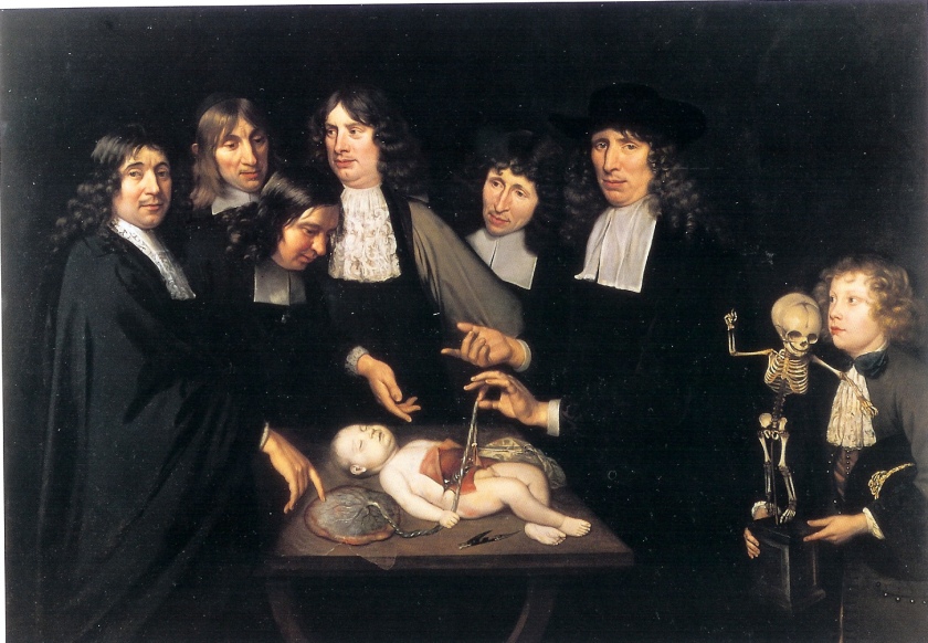
The Anatomy Lesson of Dr Frederik Ruysch; Jan van Neck 1683
Not so her brother, Hendrik. His portrait is that of a young boy, a still embryonic doctor and anatomist. In 1683 Jan van Neck paints The Anatomy Lesson of Dr Frederik Ruysch, showing members of the Surgeons’ Guild. They examine the placenta of the well-preserved body of an over-large but newborn baby. The baby is pink, apparently merely sleeping although Ruysch has opened up his abdomen. The tracery of placental blood mimics the lace cravats of the surgeons, and the umbilical cord is a gilded rope. Hendrik, at that time aged twenty, is shown as a boy and holds a baby’s skeleton.
So many babies! ‘Where do they all come from? Where do they all belong?’
Rachel and her husband, the portrait painter and lace-merchant Juriaen Pool, were to have ten children. We do not know if any of their babies died. But would she and Juriaen have asked her father to embalm them, so they might live forever? We would like to think it was unlikely, but we cannot tell.
Ruysch’s babies were not for entertainment or even to be used as specimens for teaching anatomy, they were artworks — and moralistic in tone. They were symbols of Vanitas mundi, the pointlessness of pleasure: ‘we’re doomed, we’re going to die!’ (Ruysch himself died in 1731, at the great age of 93, but it isn’t recorded whether his longevity was due to inhalation of liquor balsamicus fumes.) They were not regarded at that time with horror or disgust. Death was everywhere, a daily occurrence, it was God’s Will (a Calvinistic God, at that) so we would do well to remember the transience of our lives on earth, and the ultimate irrelevance of earthly objects. Ruysch’s babies were all perfect, and perfectly virtuous and innocent.
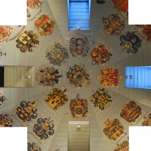
The ceiling of the Surgeons’ Guild, Der Waag. Ruysch’s shield is in the centre
Ruysch certainly had access to large numbers of both normal, and teratologically abnormal, foetuses. In 1668 he was entrusted with the training of Amsterdam’s midwives. Ten years later he was appointed as ‘doctor to the court’, which allowed him to take possession of all the dead babies found in the harbour. It was a period of history when birth rates and mortality rates were high, for many reasons. Today we are scarcely replacing ourselves, the rate of reproduction is 2.1 in Britain, even less in Italy, that country of babies and extended families; unwanted babies need no longer be conceived.
But unwanted babies will always find their way into the world, and be abandoned. A thin wail, a choking cry, comes from a telephone kiosk, a doorstep, a handbag on Paddington station. In Hamburg there is a ‘postbox’ where a desperate mother can lift the flap and leave her baby on a warm and comfortable bed, no questions asked, no identification necessary. (Is there a warning, as on Low Bridges – ‘Max width, max height’ – to prevent over-large parcels being posted?) The newly-delivered baby is lifted from the bed, kind hands stroke her silky hair and touch the pulsing fontanelle. Does she understand that comforting kiss, that it signifies Life?
Ruysch’s babies mock the frailty of their parents, who gave them brief life. A foetal hand, its severed wrist hidden by a lacy cuff, plays ball with a ‘segmentum humani testiculi’; in another jar, the lace-capped leg of a baby rudely kicks a prostitute’s syphilitic skull.
(1) From Death Enlightened (1970) A M Luyendijk-Elshout. JAMA 212, 121-126
From 2010-2011 I was a ‘Bright Ideas’ Visiting Fellow at the European Genomics Forum (EGF), University of Edinburgh. My aim was to explore and write factual or fictional accounts of some of the people whose tissues and organs came to be publicly exhibited in the Surgeons’ Hall Museum, Edinburgh.
As a contrast to these stories of past ‘donors’ I felt it was important to find out more about the way in which we now treat donation and collection of organs and tissues, for both teaching, research and transplant purposes. The stories of the people who responded are recorded in the EGF archive.
***
The great anatomy and pathology collections were initially intended as aids to teaching, but their purpose has changed with time (see the perceptive article, ‘Making sense in the pathology museum‘ by Dr Steve Sturdy, formerly EGF’s Assistant Director). The collections at the Surgeons’ Hall Museum (SHM) in Edinburgh date from the late 17th century and it is impossible not to wonder about the people – the ‘patients’ as Andrew Connell, the then Collections Manager, touchingly refers to them – whose organs and skeletons are in storage or on display.
These exhibits were ‘donated’ by human beings, each of whom had a life, perhaps in a town, on a farm, perhaps with a family and friends; or perhaps he or she was ridiculed and despised.
I had previously been shocked out of being a detached observer when I visited exhibitions and museums researching background material for my novel, The Embalmer’s Book of Recipes: at the Museum Vrolik in Amsterdam, the clatter of coffee cups being laid out for a conference, the loud talking and laughter of men repairing the heating ducts, the bright lights and chrome and glass of the display areas, suddenly clashed agonisingly with the contents of the jars – late-stage foetuses and neonates, each with some developmental or genetic abnormality. These specimens were once, briefly, sentient beings, brought into the world by their mothers, women of all ages and backgrounds, privileged or poor, who may or may not have had the sympathy and support of family or friends.
And so began my need to explore and write the stories of some of those people whose tissues and organs came to be publicly exhibited – in this case, in the Surgeons’ Hall Museum, Edinburgh.
Some of these ‘specimens’ were certainly obtained without the patient’s consent, and Andrew Connell’s musings (see “The Curator’s Reflections” ) helped me to understand why the patients might have been brought to such straits, in hope or fear or ignorance.
I was further helped in my hunt for information by Laura Brouard at the Lothian Health Services Archive in the University Library; by staff at the National Library of Scotland; and by Anne Carroll, archivist at Perth Library.
It was a special privilege to meet and spend hours talking with artist Joyce Gunn Cairns, who has drawn several of the exhibits at the Museum with great delicacy and empathy and allowed me to include her drawings and words. Poets Christine De Luca and Diana Hendry had also written about some of the exhibits – ‘Janet’ and ‘Andrew’ in particular – and to listen to them talk about their subjects was a great pleasure and gave new insights (and to hear Christine read in the Shetland dialect was an extra treat); they too have been happy for me to include their poems.
Part-way through my Fellowship, I organised a meeting that included a lawyer, a bioethicist, the curators, the poets, scientists, sociologists and writers and we had a very helpful and thoughtful discussion about my research and writing, and its ethical implications.
Finally, in February 2013, the EGF organised a well-attended ‘Tell them our stories‘ event for the public in the University’s Anatomy Theatre. The SHM’s Curator Andrew Connell and I both presented illustrated talks; poet Diana Hendry read her moving poem about ‘Andrew’, and actor Michael Flett gave an excellent and very funny reading of ‘Caesar’s story‘ (an extract from my ‘The stories of Janet and Caesar‘)
Steve Sturdy, 2006, Making sense in the Pathology Museum, in Anatomy Acts: how we come to know ourselves. Eds Dawn Kemp & Andrew Patrizio; Birlinn.
ANDREW CONNELL, Collections Manager at the Surgeons’ Hall Museum in 2011, talks about the Collection’s modern function.
“It’s like inviting people in and showing them round your house and what’s on your shelves – there are different hands exploring the collection in different ways. It brings out endless possibilities, like a kaleidoscope.”
“The first collection, which was very basic, not very good, was held in places like Infirmary Street and High School Yards. Robert Knox [1791 -1862] was made responsible for increasing the collection and for its move to new premises. He bought up Charles Bell’s collection and brought it up from London. Much of it was stored in Infirmary Street and in various corridors and cellars. Knox was also in charge of presentation and the collection was opened to the public from that time. So why would you come to see it?
“There’s a nice piece in the Tansey & Mekie ‘History of the Museum‘:
‘Visitors of the lower classes, mechanics, sailors and soldiers have uniformly been quiet, careful and most orderly…visitors of the lower classes seem to take more interest in the specimens than those of the higher, many of whom, especially ladies, merely walk through the rooms without looking at the objects particularly.’
Probably they were embarrassed to show too much interest!”
“The original context of the collection – the organs and tissues – was objective.
It was for teaching, for learning anatomy, all of which was observation-based.
For more than 150 years this was its raison d’être.
The teaching role was more a fade-out process from the 1970s/80s – that was
the start of its serious demise. With the advent of desk-top viewers for slides
you had the ability to represent material in different ways. There were other
diagnostic tools such as X-rays. And in terms of fairness in exams – you can
use the same slide for everyone rather than trying to get several identical
specimens. A photo of, for example, a pulmonary embolism is more realistic,
in its fresh natural coloured state, than a greying specimen in a jar.”
” ‘Benign neglect‘. That’s what I like to call it. We’re very happy to be a figure-
head for benign neglect. At first the collection was loved and cared for and
funded for teaching purposes. But the College is very reactionary, very slow to
follow trends, and the collection became semi-obsolete for such a long time.
But now, instead of using it to look purely at anatomy and pathology, we can
use it to look at the history of medicine, to tell a consistent tale about medicine, the medical personalities and pioneers, and also the context of the social history.”
“If we think about the patients who formed the collection – what was their life
like?
What misfortune led them here, with what stoicism?
We have to think about the things people put up with – and how they were affected.
We aim now to bring out the ‘patient-based’ context – the humanity, and
human stories.
This is where artists of all genres are important.”
***
“I started in the Chambers Street Natural History Museum, then I moved to
the University’s Department of Pathology collection. There was a certain
element of haphazardness – I liked working with my hands, and enjoyed fixing
things. Chambers Street was quite Dickensian, with the inventories still on
cards. At the University it was more hands-on, a very different collection – I
had workshop duties in anatomy and pathology, teaching duties such as
display sets for classes, and I repaired things.
Medical terminology? I made an effort, enough to get by on – but it was
‘shallow waters’. Don’t take my word for anything – you should demand a
second opinion!”
“When I was in Pathology, Dr Black once handed me a poly bag with a kidney
in it, still warm. He just said, ‘Can you deal with this?’ There was a bit of a
confrontation. ‘Am I supposed to?’
For me, the first issue was mortality – this was somebody’s organ, used until not long ago. Yes, the kidney was ‘dealt with’, but I then came away from that experience with some issues.
In the Museum, it isn’t red, it isn’t warm – and you start becoming aware of
your own feelings in relation to these aspects.”
“The society we live in, we’ve become desensitised to death, it’s all neatly
packaged in boxes. So you don’t know how somebody will react to a museum
specimen. When people come round, I’m very conscious of their response to
the collection. I try to think, what’s right, what’s wrong, about having that
specimen on show? But the responses are deeply personal, a gut reaction,
intuitive.
I might be overly protective, overly cautious, of how we present things to people.
For example, we had Siamese twins – they were on display in an educational
context, with X-rays that showed how their bones were joined. But then I used
to hear people coming in and saying, ‘Where’s the babies in jars?’ They were
missing the point, it was just voyeuristic, they were treating it like a Victorian
horror-show.”
***
“I want to get people to look without feeling distressed about mortality: to start
a reflective process that’s speculative – and they can then allow themselves to
go in different directions. I want people to use the collection as a locus for opening up, and contextualising in the way of social history.
There’s an inter-connectedness between people – the surgeons, patients,
pathologists, the family – and they’re all colliding and ricocheting in all directions.
The collection here provides the impetus for you to look differently, and to re-focus your thoughts and ideas.”
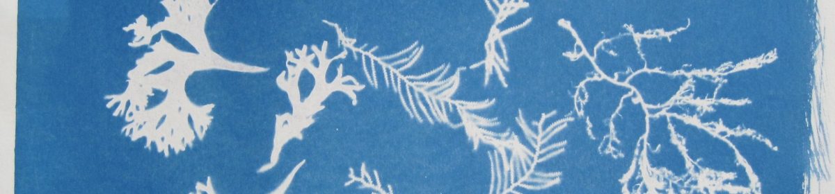


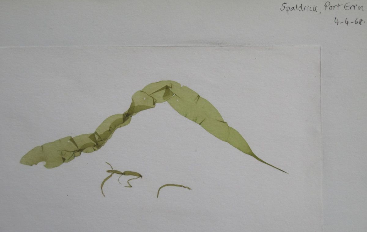


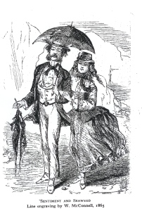


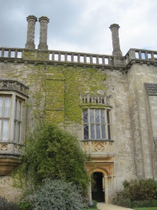


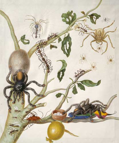
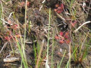


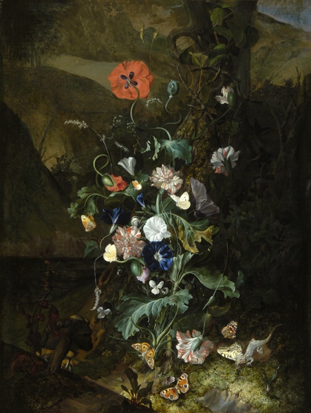


")